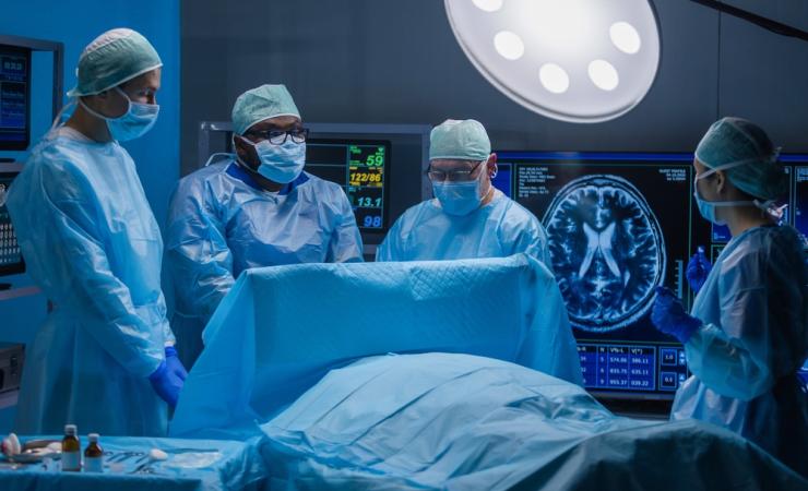
In many fields, surgeons are increasingly using minimally-invasive techniques to carry out operations – incisions are kept to a minimum, and surgeons are guided in their work by imaging technologies that show what is happening inside the patient in real time. In contrast, neurosurgery still typically entails making large openings in the skull, and attempts to integrate imaging technology into neurosurgery procedures are hampered by costs and logistics. As a result, patients with many neurological conditions are still undergoing operations where the risk of complications (such as brain damage, infections and consequential poor cosmetic outcomes and decreased quality of life) is high.
Brain surgery “one of the greatest challenges in modern medicine”
“The brain is extraordinarily complex and delicate. It defines who we are, including our intelligence, personality, and consciousness,” said SEISMIC academic lead Pieter Kruizinga of Erasmus MC. “Operating on the brain remains one of the greatest challenges in modern medicine, as the margin between helping and harming a patient can be extremely small. SEISMIC is about shifting that balance in the right direction by making neurosurgical procedures less invasive, more precise, and better supported by real-time imaging, data, and training. By bringing these elements together, we aim to improve outcomes for patients and enhance the way neurosurgeons work.”
SEISMIC aims to improve patient care by working out how to seamlessly integrate medical imaging technologies into minimally invasive neurosurgery techniques. The team will focus on three conditions where surgery is essential for patient survival and quality of life: intracerebral haemorrhage (ICH, bleedings deep in the brain), chronic subdural haematoma (cSDH, bleedings between the skull and the brain) and brain tumours (where surgery is key during both diagnosis and treatment).
Stopping bleeding the SEISMIC way
On ICH, SEISMIC will explore how imaging techniques could be used to guide a small drainage device directly to a bleed deep in the brain via a small hole in the skull, allowing the haemorrhage to be drained more accurately and with a lower risk of damage to healthy brain tissue.
In the case of subdural haematomas, the team will investigate how to combine in one procedure drainage of the bleed with blocking the flow of the middle meningeal artery, all supported by imaging technologies. This is important because in 30% of patients, the bleeding starts again following surgery, and blocking the middle meningeal artery could prevent this from happening.
Better diagnosis and treatment of brain tumours
Today, diagnosing a brain tumour often requires brain surgery to obtain a sample of the tumour for analysis. To reduce the need for surgery, SEISMIC plans to see if it would be possible to get diagnostic information on brain tumours from a blood sample. The challenge here is that thanks to the blood-brain barrier (BBB), brain tumour markers rarely get into the blood. Research indicates that injecting microbubbles and applying focused ultrasound (FUS) can locally activate the bubbles, temporarily opening the blood–brain barrier (BBB). This may enable tumour markers to enter the bloodstream, making them easier to sample.
Finally, the team will assess the use of cryoablation (i.e. the use of extreme cold) to destroy brain tumours. Cryoablation is already used to treat other cancers, but has not yet been applied to brain tumours, which are typically removed via open brain surgery. The cryoablation device would be inserted via a small hole in the skull and guided to the tumour using imaging techniques such as X-rays and ultrasound.
All four minimally invasive procedures will be tested in clinical trials. In addition to evaluating the effectiveness of the procedures, the project will also assess their operational efficiency, economic viability, and scalability in real-world settings.
Barriers to the wider use of imaging during brain surgery include the sheer size of imaging equipment, lengthy set-up times that block the operating theatre, and the need for extensive training in their use. To achieve its goals in all three disease areas, SEISMIC will set out to deliver ground-breaking innovations in both ultrasound and X-ray imaging technologies, as well as systems to allow their seamless integration in neurosurgery.
By integrating imaging technology and minimally-invasive procedures for neurosurgery, SEISMIC could help to dramatically cut the need for open surgery for people with intracerebral haemorrhage, chronic subdural haematoma, and brain tumours. This would improve patients’ lives by improving outcomes, cutting recovery times, and reducing the risk of complications.
“Tremendous potential to improve patient care”
“The shift from open brain surgery to minimally invasive procedures, supported by advanced imaging and precision navigation, holds tremendous potential to improve patient care,” said Bert van Meurs of Royal Philips. “It’s a major undertaking – and through SEISMIC, we’re bringing together the necessary medical specialties, technology domains and patient perspectives to help break down the barriers to this transition. By combining precision surgical techniques with real-time imaging and intelligent decision support, our goal is to simplify complex procedures, improve outcomes and broaden access to advanced neurosurgical care across Europe and beyond.”