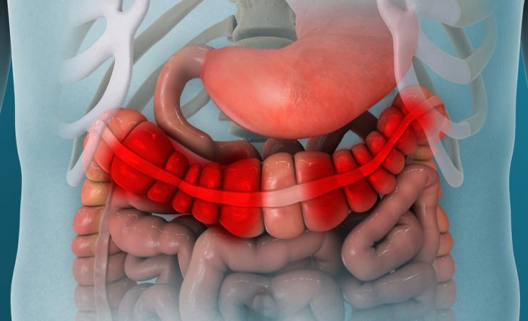
Inflammatory bowel diseases (IBD) can happen when the intestines are inflamed, resulting in poor digestion and gastro-intestinal issues. Crohn’s disease and ulcerative colitis both belong to this group of disorders. There is no cure for IBD, and almost five million people worldwide have these diseases, which cause them to experience painful flare-ups for the rest of their lives.
Treating IBD is complex – what works for one patient won’t necessarily work for another. Vedolizumab, a type of monoclonal antibody, works as a treatment for about half of all IBD patients, but there is as yet no way to determine which patients will respond well to this treatment. The way that the drug works inside the body is not well understood, so the IMI Immune-Image project used a technique called fluorescence molecular imaging to track the journey of vedolizumab through the body.
“The main goal of the research was to see where vedolizumab goes in the body and which cells it affects. We did this by developing fluorescence vedolizumab that lights up in the bowel of the patients with IBD,” says Anne van der Waaij of the University of Groningen, one of the authors of the paper.
By attaching fluorescent dye to vedolizumab before giving it to patients, the researchers could track the progress of the drug and see which targets it bound to. The project carried out forty-three fluorescence imaging procedures in thirty-seven patients with IBD.
“Thanks to the funding we’ve received from the IMI partnership, we showed that fluorescence imaging enables the visualisation of fluorescently labelled medication in the human bowel during endoscopies and biopsies. We also saw increased signals when the dose of fluorescently labelled vedolizumab was increased,” says Ruben Gabriels of the University of Groningen, the first author of the study.
They saw that the drug targets a specific variety of immune cell types within the inflamed tissue – a group that were previously not suspected of being involved.
“Fluorescence microscopy showed that vedolizumab is linked to different types of immune cells than previously thought, giving us new ideas about how the drug works,” says Pia Volkmer of the University of Groningen, who was also involved in the study.
This was the first time that imaging techniques were used to map the journey of vedolizumab through the body, illustrating how imaging technology can help to better inform pharmaceutical development.
The hope is that these results might lead to a better understanding of how the drug functions which could eventually help predict which patients would benefit from using it, as well as highlighting who might need a new drug and why.
The next steps for the Immune Image project will be to predict the body’s response based on fluorescence signal intensity, and to compare different kinds of IBD treatment.
Immune-Image is supported by the Innovative Medicines Initiative, a partnership between the European Union and the European pharmaceutical industry.
