In coronary artery disease (CAD), plaques made up of fat and calcium build up in the arteries, gradually narrowing them and blocking the flow of blood to the heart. It affects around 5% of people in the EU and is a leading cause of death.
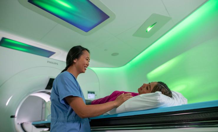
According to current clinical guidelines in the EU, UK and US, patients suspected of having CAD should first have a scan called a coronary computed tomography angiography (CCTA). CCTA is non-invasive and uses X-rays and computer processing to create a detailed 3D image of the coronary arteries. It therefore allows doctors to accurately assess how blocked a patient’s arteries are and helps to determine whether the patient needs an invasive procedure such as an operation to manage their condition or if medication would be enough.
In practice, challenges with interoperability across systems, devices, and geographies mean that CCTA is not being used to its full potential. As a result, an estimated 60% of patients still undergo invasive tests such as invasive coronary angiography (ICA) for CAD unnecessarily. In an ICA, a catheter is inserted into the blood vessels at the groin or arm and guided to the heart area where it releases a contrast medium that shows up on X-rays.
The goal of the COMBINE-CT project is to deliver an automated CCTA-enabled workflow that will improve and connect the different steps in the care of CAD patients, ranging from diagnosis to treatment planning, treatment procedure, and follow-up. Artificial intelligence (AI) powered algorithms will make it easier than ever to definitively diagnose CAD, select the right treatment for the right patient, plan interventions, use this information during an invasive procedure, and ensure patients are followed up appropriately.
“With the COMBINE-CT project we want to improve the care pathway for coronary artery disease, the most common heart disease,” said Bert van Meurs, Chief Business Leader Precision Diagnosis and Image Guided Therapy at Philips. “The introduction of new technology creates more data that we can use for diagnosis and treatment. Due to interoperability challenges this information is not used to its full potential. In COMBINE-CT, we will work with top clinical centres and industry partners to open the data silos in the hospitals between different departments involved in the care of CAD patients, simplifying, and improving workflows for physicians, nurses, and technologists.”
The project brings together experts in cardiology, imaging, surgery, and artificial intelligence as well as health economists and patient organisations. Together, they will carry out five multi-centre studies to generate evidence on the benefits of computed tomography (CT) scans across the CAD care pathway. Among other things, these studies will investigate the value of CT for definitive CAD diagnosis, treatment planning and support, and explore how a technique called photon counting spectral CT can be used to evaluate coronary artery bypass grafts and previously implanted stents.
“The COMBINE-CT project will have significant impact on clinical practice by leveraging the power of artificial intelligence combined with CCTA, and facilitating closer collaboration between radiologists and cardiologists to optimise the diagnostic and therapeutic pathway for patients with suspected CAD,” said Dr Bimmer Claessen, interventional cardiologist at the Amsterdam University Medical Centres. “This will lead to a reduction in unnecessary invasive coronary angiography procedures. Moreover, it will allow careful pre-procedural counselling of patients with complex coronary artery disease, meticulous pre-procedural planning, and procedural guidance for percutaneous coronary interventions. Additionally, we are investigating the use of CCTA biomarkers as a means to evaluate the effectiveness of medical therapy aimed at reducing the progression of atherosclerosis in diabetic patients.”
While COMBINE-CT focuses on heart disease, the project hopes that its findings will also prove useful to other disease areas where CT scans are widely used.
